Ventricular Septal Defect: Abnormal Blood Flow
 |
| A ventricular septal defect (VSD) is an opening in the interventricular septum, causing a shunt between ventricles. Large defects result in a significant left-to-right shunt and cause dyspnea with feeding and poor growth during infancy (Beerman, 2023). |
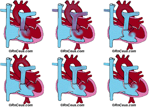
Ventricular septal defect (VSD) is classified as an "acyanotic" congenital heart defect. In an acyanotic condition, the abnormal blood flow pattern occurs without introducing significant volume of deoxygenated blood (blue) into the systemic arterial circulation. The animation on the right side of the page, in part, demonstrates oxygenated blood (red) being shunted by higher pressure from the left ventricle through a large septal defect where it combines with the deoxygenated blood in the right ventricle. During systole the combined blood volume is then forced through the pulmonary valve and into the pulmonary artery to be recirculated through the lungs. This characterizes an acyanotic VSD.
Large VSDs, like the one depicted here, increase both the volume and pressure of blood entering the pulmonary vasculature. It is important to note that an infant with a large VSD may be asymptomatic in the first few days/weeks of life until the pulmonary system fully expands, matures and reaches its ability to accomodate. Left untreated, the increased blood flow can result in pulmonary edema, causing the neonate/child to exhibit symptoms of:
Our animated example of the large VSD is further complicated by its proximity to the aorta (subaortic VSD), where in this case a small amount of deoxygenated (blue) blood is seen to flow from the right ventricle to the left ventricle through the defect during left ventricular diastole (filling) due to transient pressure differences. This can result in a small, usually inconsequential amount of deoxygenated blood circulating through the systemic arterial system.
As we will see later, this is substantially different from the significant right-to-left shunt characterized by the Eisenmenger Syndrome. Eisenmenger Syndrome can develop when a condition like pulmonary arterial hypertension increases right ventricular outflow resistance, causing blood to be shunted from right to left ventricle through the VSD.
Consequences of the Abnormal Blood Flow
Pulmonary vascular resistance is typically low, allowing the lungs to accommodate the normal blood flow. However, the additional blood volume caused by a significant left to right ventricular shunt can lead to:
It's important to note that the size of the VSD significantly influences the magnitude of the shunt and the severity of these consequences. Smaller VSDs may result in minimal hemodynamic effects, while larger VSDs can lead to significant volume overload and early development of heart failure.
References
Cleveland Clinic. (2025). Ventricular Septal Defect (VSD). Accessed 4/12/2025 from https://my.clevelandclinic.org/health/diseases/17615-ventricular-septal-defects-vsd
Beerman, L.B. (2023). Ventricular Septal Defect. Merck Manual, Professional Version. Accessed 4/12/2025 from https://www.merckmanuals.com/professional/pediatrics/congenital-cardiovascular-anomalies/ventricular-septal-defect-vsd
© RnCeus.com